This example will give you some insights about Cell Tracking Challenge (CTC) data on CellTrackVis.
1. BF-C2DL-HSC
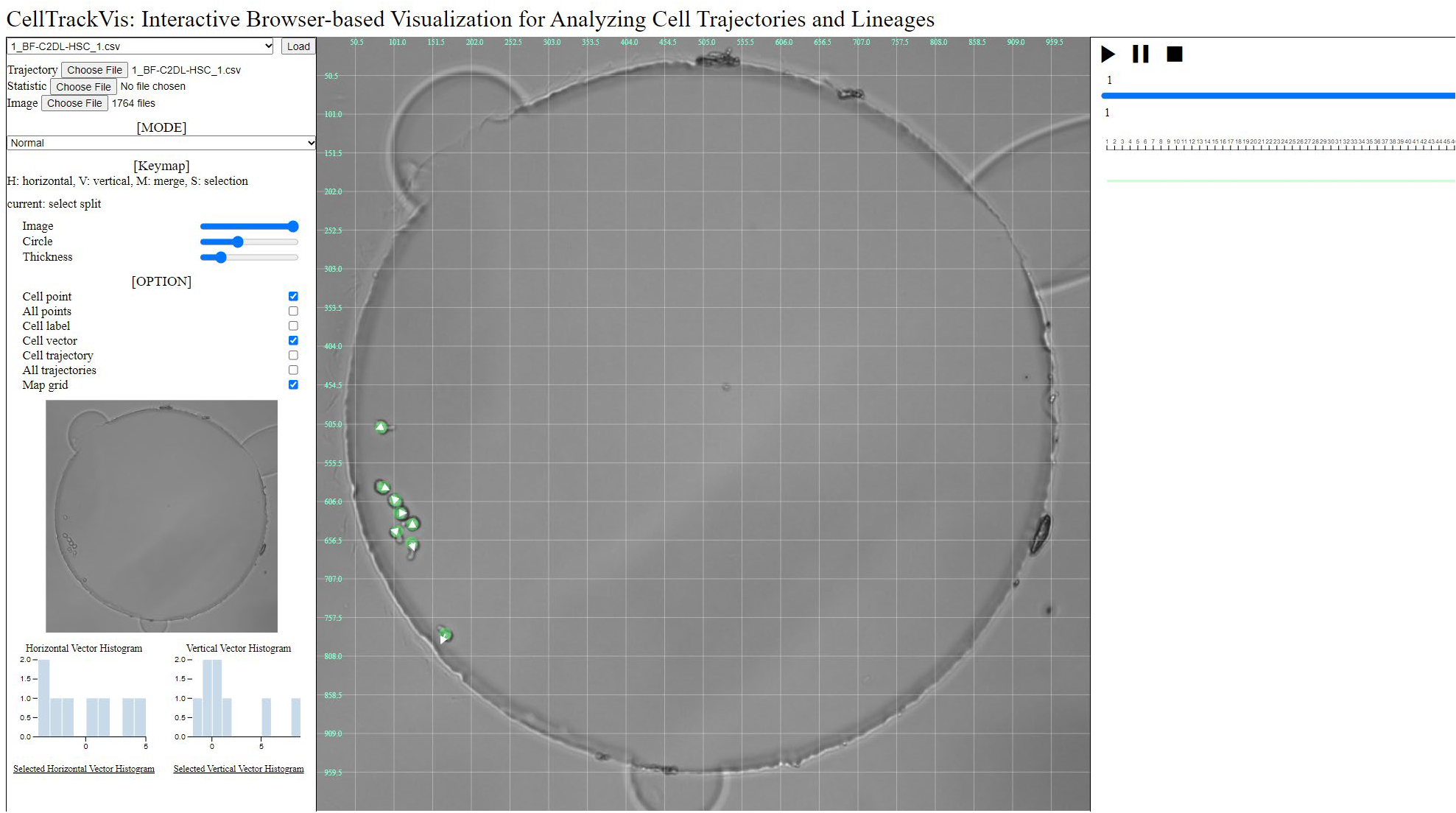
A ground truth of the long image sequences in CTC is plotted using CellTrackVis.
Mouse hematopoietic stem cells in hydrogel microwells
Dr. H. Blau, Baxter Laboratory for Stem Cell Biology, Stanford University, USA

Microscope: Zeiss PALM/AxioObserver Z1
Objective lens: EC Plan-Neofluar 10x/0.30 Ph1
Pixel size (microns): 0.645 x 0.645
Time step (min): 5
2. BF-C2DL-MuSC
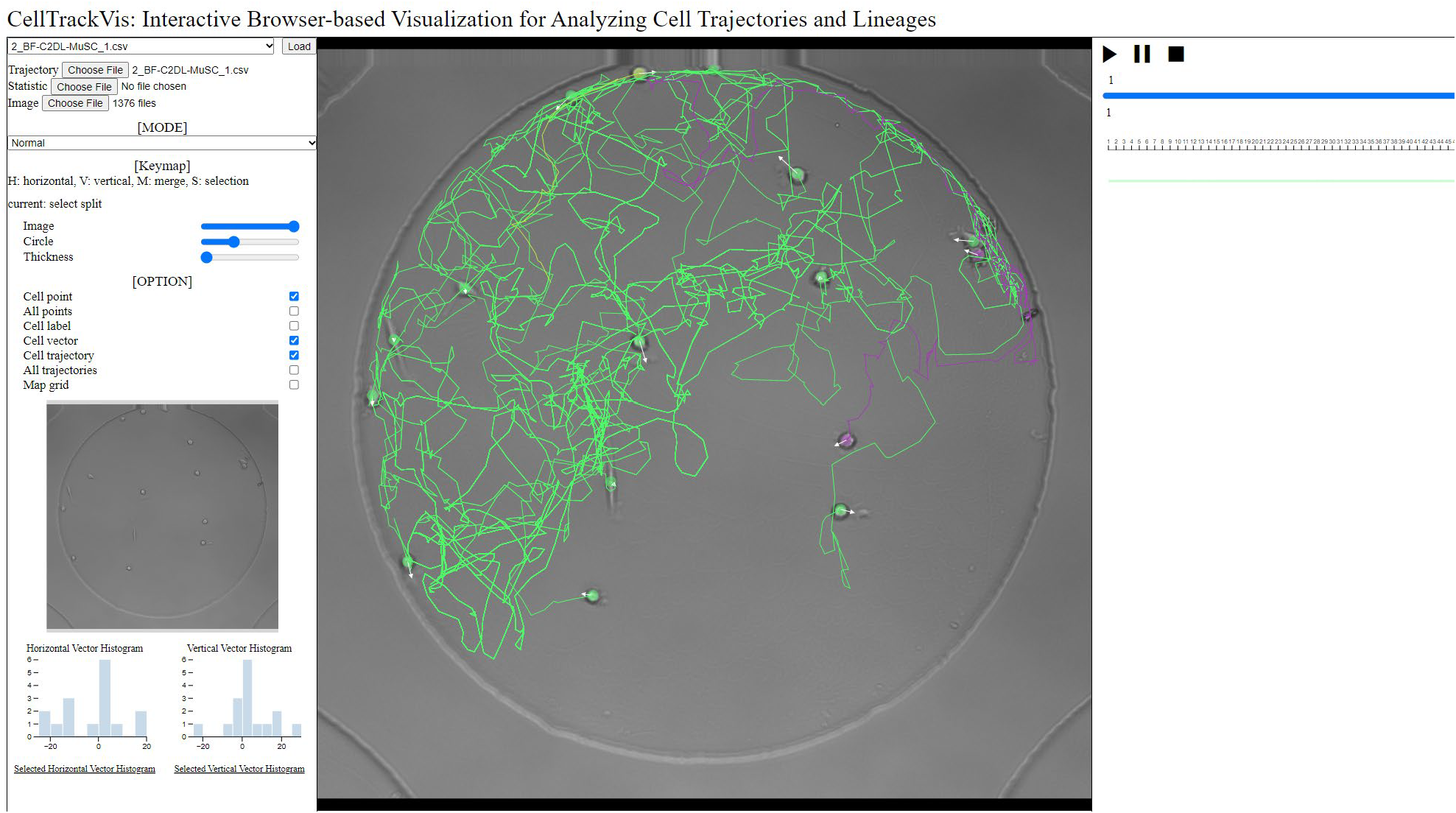
A ground truth of the long image sequences in CTC is plotted using CellTrackVis.
Mouse muscle stem cells in hydrogel microwells
Dr. H. Blau, Baxter Laboratory for Stem Cell Biology, Stanford University, USA

Microscope: Zeiss PALM/AxioObserver Z1
Objective lens: EC Plan-Neofluar 10x/0.30 Ph1
Pixel size (microns): 0.645 x 0.645
Time step (min): 5
3. DIC-C2DH-HeLa
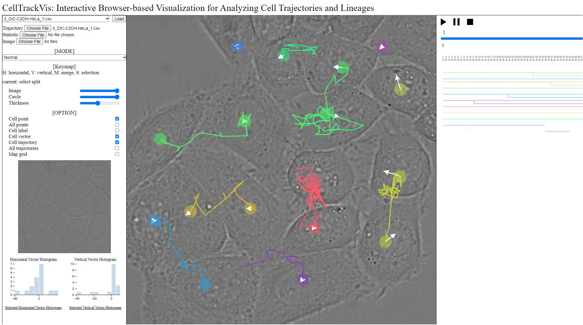
A ground truth in CTC is plotted using CellTrackVis.
HeLa cells on a flat glass
Dr. G. van Cappellen. Erasmus Medical Center, Rotterdam, The Netherlands

Microscope: Zeiss LSM 510 Meta
Objective lens: Plan-Apochromat 63x/1.4 (oil)
Pixel size (microns): 0.19 x 0.19
Time step (min): 10
4. Fluo-C2DL-Huh7
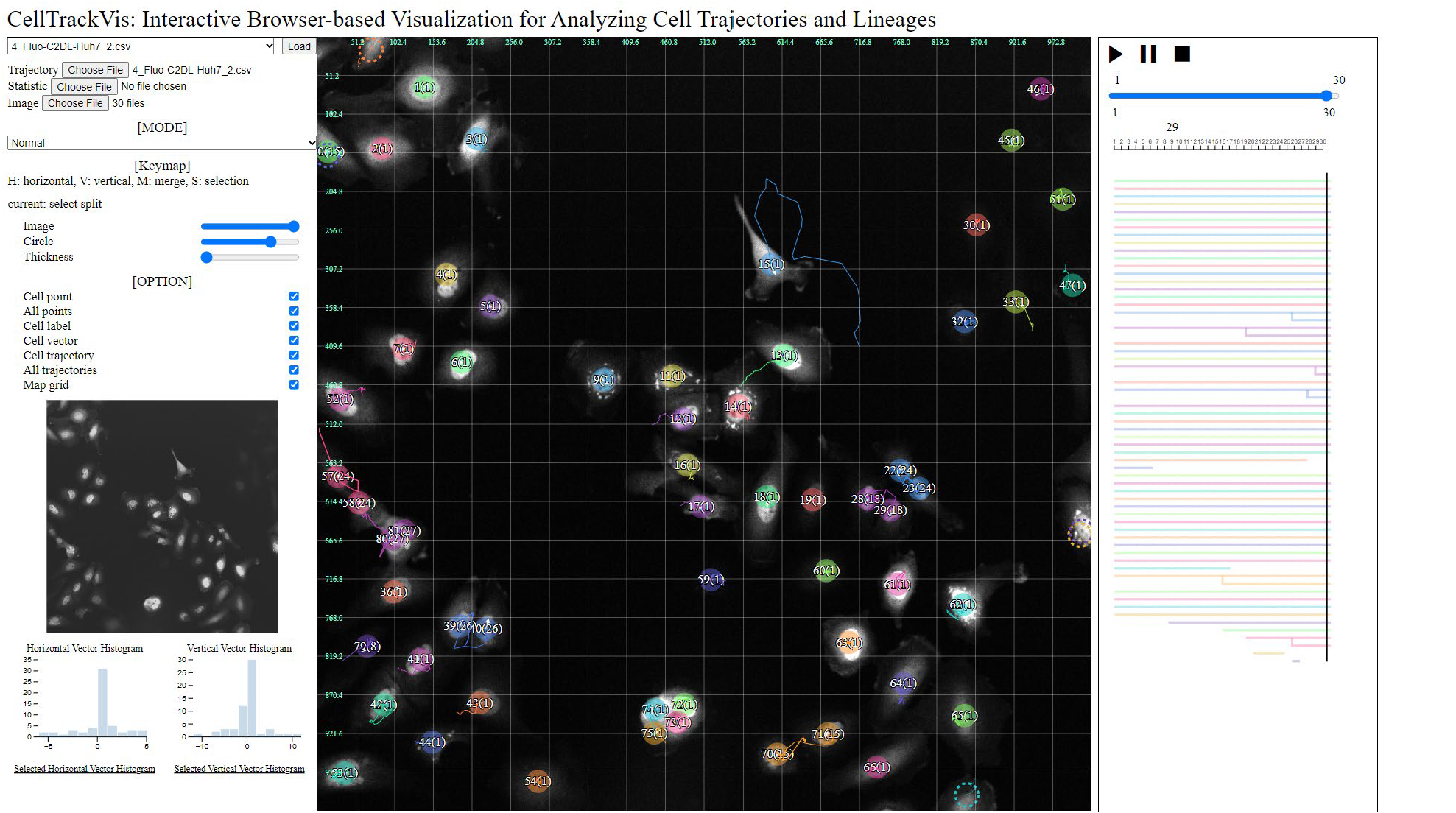
A ground truth in CTC is plotted using CellTrackVis.
Human hepatocarcinoma-derived cells expressing the fusion protein YFP-TIA-1
Dr. Alessia Ruggieri and Philipp Klein, Centre for Integrative Infectious Disease Research (CIID), University Hospital Heidelberg, Germany

Microscope: Nikon Eclipse Ti2
Objective lens: CFI Plan Apo Lambda 20x/0.75
Pixel size (microns): 0.65 x 0.65
Time step (min): 15
5. Fluo-C2DL-MSC
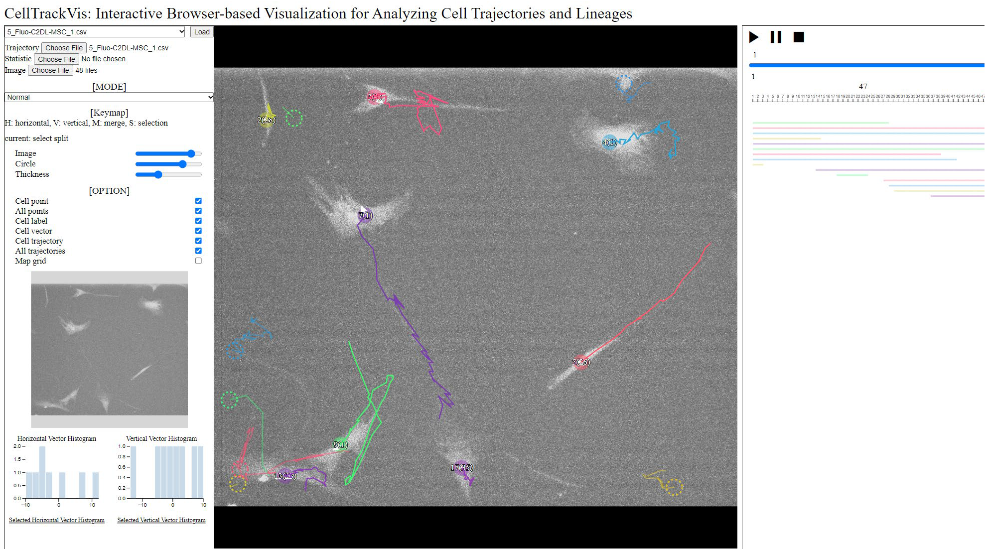
A ground truth in CTC is plotted using CellTrackVis.
Rat mesenchymal stem cells on a flat polyacrylamide substrate
Dr. F. Prósper. Cell Therapy laboratory, Center for Applied Medical Research (CIMA), Pamplona, Spain

Microscope: PerkinElmer UltraVIEW ERS
Objective lens: Plan-Neofluar 10x/0.3 (Plan-Apo 20x/0.75)
Pixel size (microns): 0.3 x 0.3 (0.3977 x 0.3977)
Time step (min): 20 (30)
6. Fluo-N2DH-GOWT1
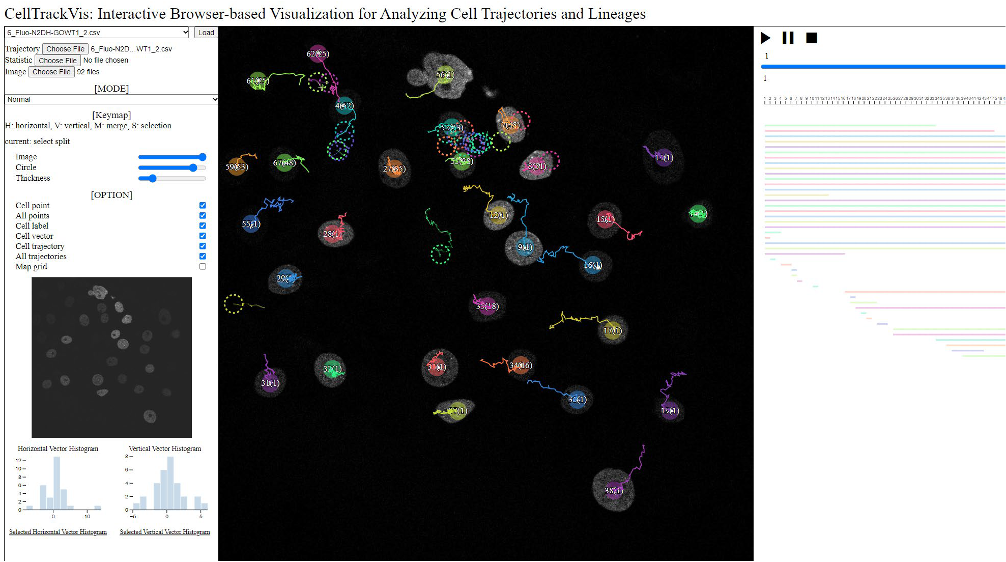
A ground truth in CTC is plotted using CellTrackVis.
GFP-GOWT1 mouse stem cells
Dr. E. Bártová. Institute of Biophysics, Academy of Sciences of the Czech Republic, Brno, Czech Republic

Microscope: Leica TCS SP5
Objective lens: Plan-Apochromat 63x/1.4 (oil)
Pixel size (microns): 0.240 x 0.240
Time step (min): 5
7. Fluo-N2DL-HeLa
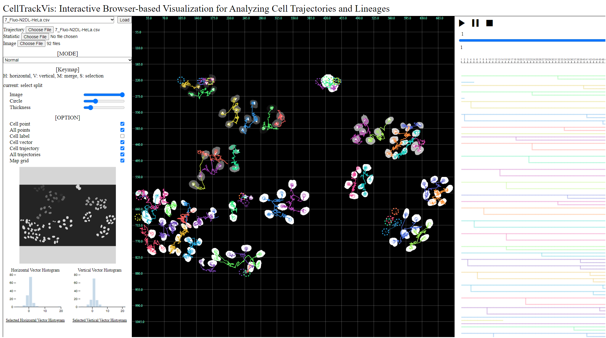
A ground truth in CTC is plotted using CellTrackVis.
HeLa cells stably expressing H2b-GFP
Mitocheck Consortium

Microscope: Olympus IX81
Objective lens: Plan 10x/0.4
Pixel size (microns): 0.645 x 0.645
Time step (min): 30
8. PhC-C2DH-U373
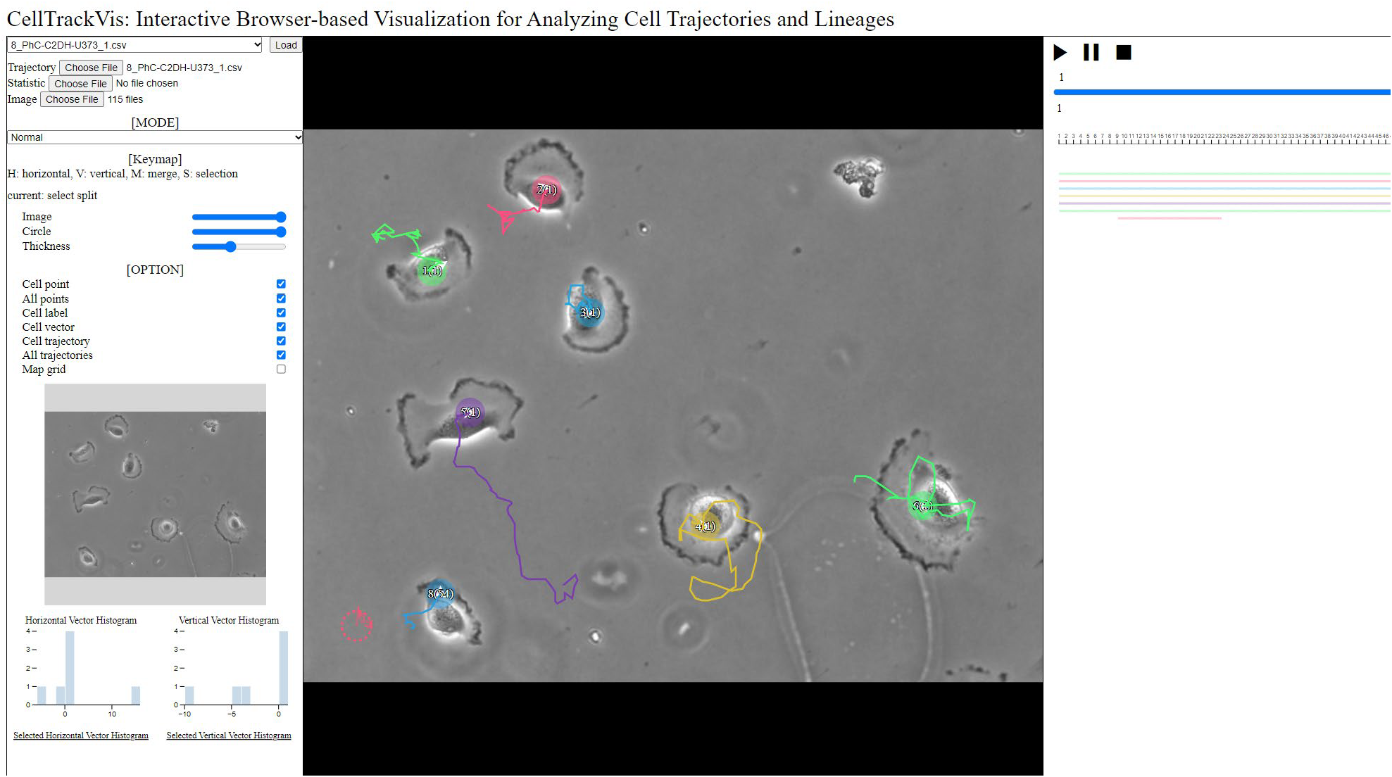
A ground truth in CTC is plotted using CellTrackVis.
Glioblastoma-astrocytoma U373 cells on a polyacrylamide substrate
Dr. S. Kumar. Department of Bioengineering, University of California at Berkeley, Berkeley CA (USA)

Microscope: Nikon
Objective lens: Plan Fluor DLL 20x/0.5
Pixel size (microns): 0.65 x 0.65
Time step (min): 15
9. PhC-C2DL-PSC
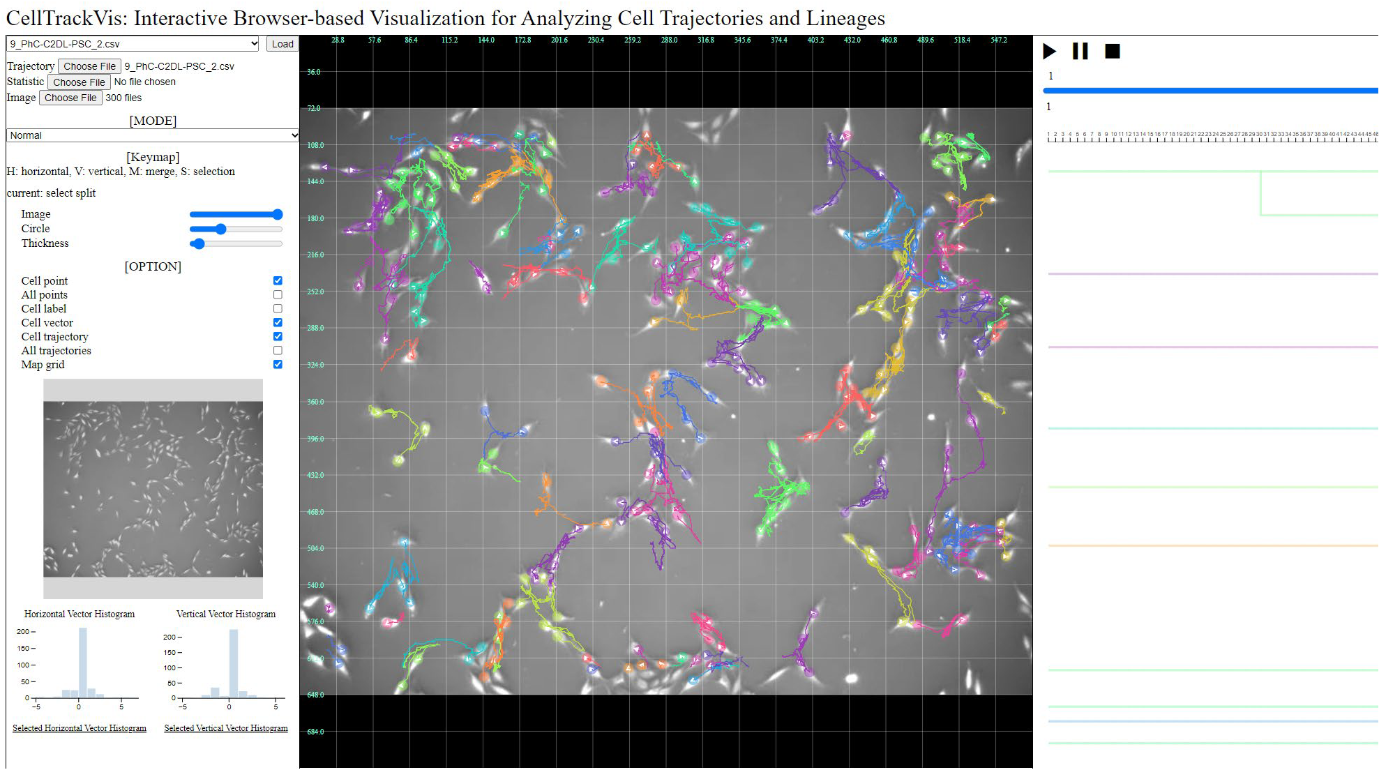
A ground truth of the large number of cells in CTC is plotted on trajectory view.
Pancreatic stem cells on a polystyrene substrate
Dr. T. Becker and Dr. D. Rapoport. Fraunhofer Institution for Marine Biotechnology, Lübeck, Germany

Microscope: Olympus ix-81
Objective lens: UPLFLN 4XPH
Pixel size (microns): 1.6 x 1.6
Time step (min): 10
10. Fluo-N2DH-SIM+
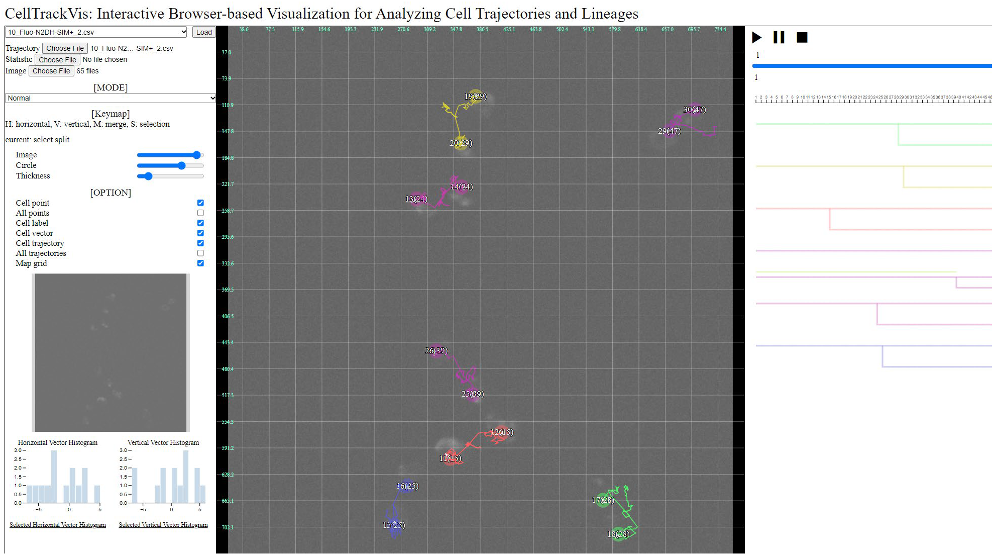
A ground truth in CTC is plotted using CellTrackVis.
Simulated nuclei of HL60 cells stained with Hoescht
Dr. V. Ulman and Dr. D. Svoboda. Centre for Biomedical Image Analysis (CBIA), Masaryk University, Brno, Czech Republic (Created using MitoGen, part of Cytopacq)

Microscope: Zeiss Axiovert 100S with a Micromax 1300-YHS camera
Objective lens: Plan-Apochromat 40x/1.3 (oil)
Pixel size (microns): 0.125 x 0.125
Time step (min): 29
Note: Visit CTC site - 2D data for further details